
INTRODUCTION
The goal of bedside renal ultrasonography is to rapidly evaluate the patient presenting to the ED with flank pain, abdominal pain with hematuria or decreased urinary output to answer a few basic questions:
Is there hydronephrosis?
Unilateral or bilateral?
Is there fluid around the kidney?
Is the bladder distended?
Are stones seen?
Is the aorta normal?
THE BASICS


INTRODUCTORY SONOSITE RENAL VIDEOS
Point of care ultrasound for renal colic will be a more focused assessment of the kidney and our main area of interest will be the collecting system.
Unlike assessing the RUQ where we are looking for a hyperechoic stone with posterior shadowing, our goal with renal US is to identify indirect signs of ureterolithiasis such as hydronephrosis.
Probe: Low frequency curvilinear probe to allow for penetration deep into tissue for visualization of the entire kidneys and bladder. If you are having difficulties with significant rib shadowing, consider the phased-array probe for a smaller footprint allowing you to squeeze between th rib spaces.
Positioning: Having the patient lay supine is a good place to start as it will allow you to scan both kidneys and the bladder fairly quickly. You can place the patient in alternating lateral decubitus positions if you are not obtaining adequate views of the kidneys supine which is often due to a large body habitus.
Where to Scan: Your scanning positions will be very similar to your FAST, so start as if you were performing this scan, placing the probe with marker towards the patient's head to obtain a long axis view of each kidney. You can try the short axis view as well by rotating the probe 90 degrees, but often the long axis view will be adequate for your evaluation.
RUQ: Mid axillary line with the middle of the probe over the costal margin, using the liver as your acoustic window.
LUQ: Start mid axillary line with middle of probe over the costal margin, then move a little more superior and posterior and you should find your kidney here. Use the spleen as your acoustic window.
HYDRONEPHROSIS (5MIN SONO)
Grading of Hydronephrosis

Hydronephrosis:
You will be looking for distention of the collecting system. In a normal kidney the renal pelvis may be minimally visible within the surrounding hyperechoic renal sinus (fat content makes it bright).
As obstruction of the ureter occurs, the renal pelvis becomes progressively dilated, leading to enlargement of the calyces and finally thinning of the renal cortex. This can be graded as as mild, moderate or severe and is quite subjective.
The above diagram gives you an idea of what you may expect to see on ultrasound when obstruction is present and a general visual grading guideline. Mild hydronephrosis will be difficult to pick up so it is essential to compare it to the opposite side and make sure you are not just appreciating a well hydrated patient.


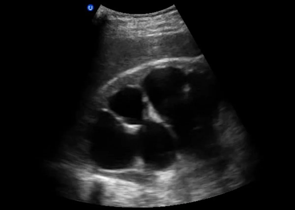
Severe Hydronephrosis
BLADDER VOLUME MEASUREMENT (5MIN SONO)
Ureteral Jets:
Assessment of ureterovesicular jet dynamics, including velocity and how frequently they are occurring, as well as asymmetry, has a pretty good sensitivity for assessing for obstruction however this takes time and in the ED may not be that useful. If you are going to take a look, place the probe over a full bladder in a transverse orientation. You will be looking posteriorly at the UVJs. See references for more information on this topic.
The Twinkling Artifact:
As we mentioned earlier, most ureteral stones will not be visualized directly on your bedside ultrasound, however color doppler may be useful to identify an obstructing stone that is initially invisible to your eye. You will be looking for the twinkling artifact, which has a 100% specificity for an obstructing stone! Note that you can assess for this both when you are performing ultrasound of the kidney as well as when you are assessing for ureteral jets (as the most common location of obstructing stone is at UVJ).
CONCLUSION
Renal ultrasound is better at identifying indirect signs of ureterolithiasis such as hydronephrosis, and not stone size and location, however we know that the majority of stones are initially managed with medical expulsion therapy.
Ultrasound of the renal system is also fast and can be performed at the bedside efficiently and accurately. With this in mind, it is reasonable to start with ultrasound for evaluation of renal colic as long as there are not complicating factors such as infected urine, severe pain or concern for more life threatening alternative diagnosis (be wary in older patients without h/o kidney stones or with multiple co-morbidities).
A few more pearls:
In an elderly patient with acute flank pain, don't forget to perform a simultaneous exam of the aorta as this is a cannot miss diagnosis and may present similarly to renal colic
A renal cyst can appear as a anechoic structure within the kidney and mimic hydronephrosis. These do not originate from the renal pelvis and usually exist isolated in the renal parenchyma.
If something looks abnormal or if you are unable to discern the architecture of the kidney well, do not ignore it, work up further with more advanced imaging
CONTINUED LEARNING
Renal Ultrasound Emergency Ultrasound Teaching
Ultrasound Vs CT for Renal Colic The SGEM